

General Information

Optical microscopy is a technique that allows the observation of mostly translucent samples, or in the case of opaque samples, with a reflection surface that is not perfectly polished. The light strikes the sample at different depths, generating a blurred image due to the detection of light from areas outside the plane of focus, which causes a significant degradation in the sharpness, contrast and resolution of the image.

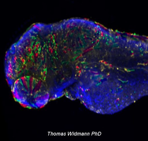
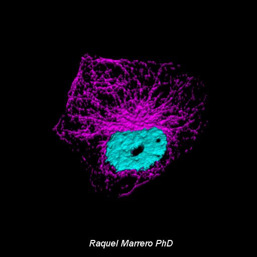
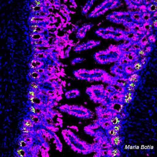
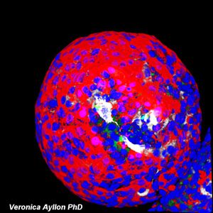
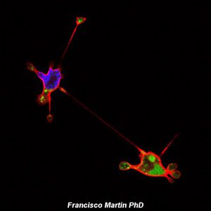
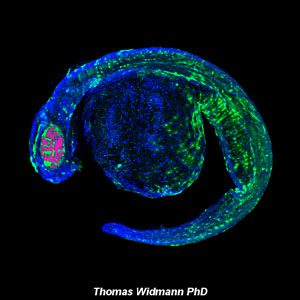
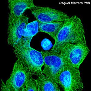
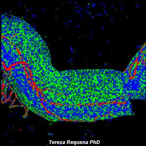
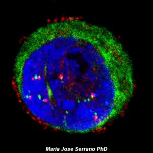
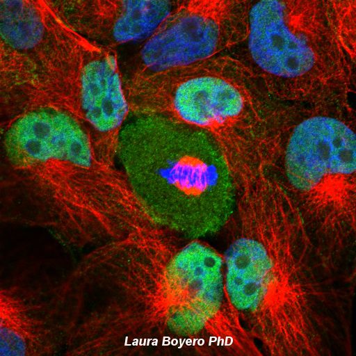
Confocal laser microscopy appeared with great success after the development of the laser, achieving excellent results in various disciplines (medicine, biology, geology, etc.), due to its undoubted advantages over traditional optical microscopy. The principle of confocal laser microscopy is based on the elimination of reflected light or fluorescence from out-of-focus planes. For this purpose, the sample is illuminated point by point with a laser line and only the light coming from the focal plane is detected, eliminating the beams from the lower and upper planes, thus generating images of high sharpness, contrast and resolution. In addition, with confocal laser microscopy, optical sections of the sample can be obtained, allowing for a three-dimensional study.

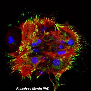
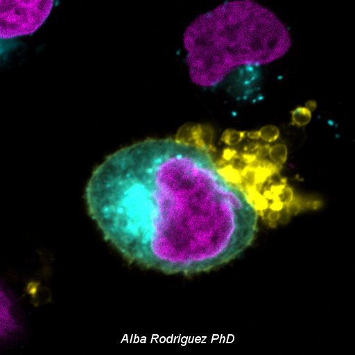
Total Internal Reflection Fluorescence (TIRF) microscopy is based on a type of illumination that generates an evanescent wave or field approximately 100 nm thick, producing excitation of a limited region of the sample, located adjacent to the interface between two media with different refractive indices. In practice, the interface most commonly used in TIRF is the contact area between the basal plasma membrane of the cell and the glass or substrate to which the cell adheres. TIRF microscopy allows a wide range of applications in cell biology, such as analysing the localisation and dynamic distribution of fluorescent molecules near the basement plasma membrane of cells.

The laser capture microdissection technique focuses on the observation and separation of single cells or small cell groups from heterogeneous tissue in a selective, efficient and rapid manner, enabling the systematic study of the quality and quantity of biomolecules (DNA, RNA or proteins) in specific cell populations. This technology, which was originally developed for the molecular analysis of tumours, can now be applied to a wide variety of tissues, and facilitates the study of the molecular basis of numerous diseases, such as those associated with human genetic variability, such as cancer, rare diseases, degenerative diseases, diabetes and hypertension, among others.

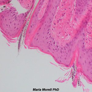
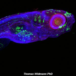
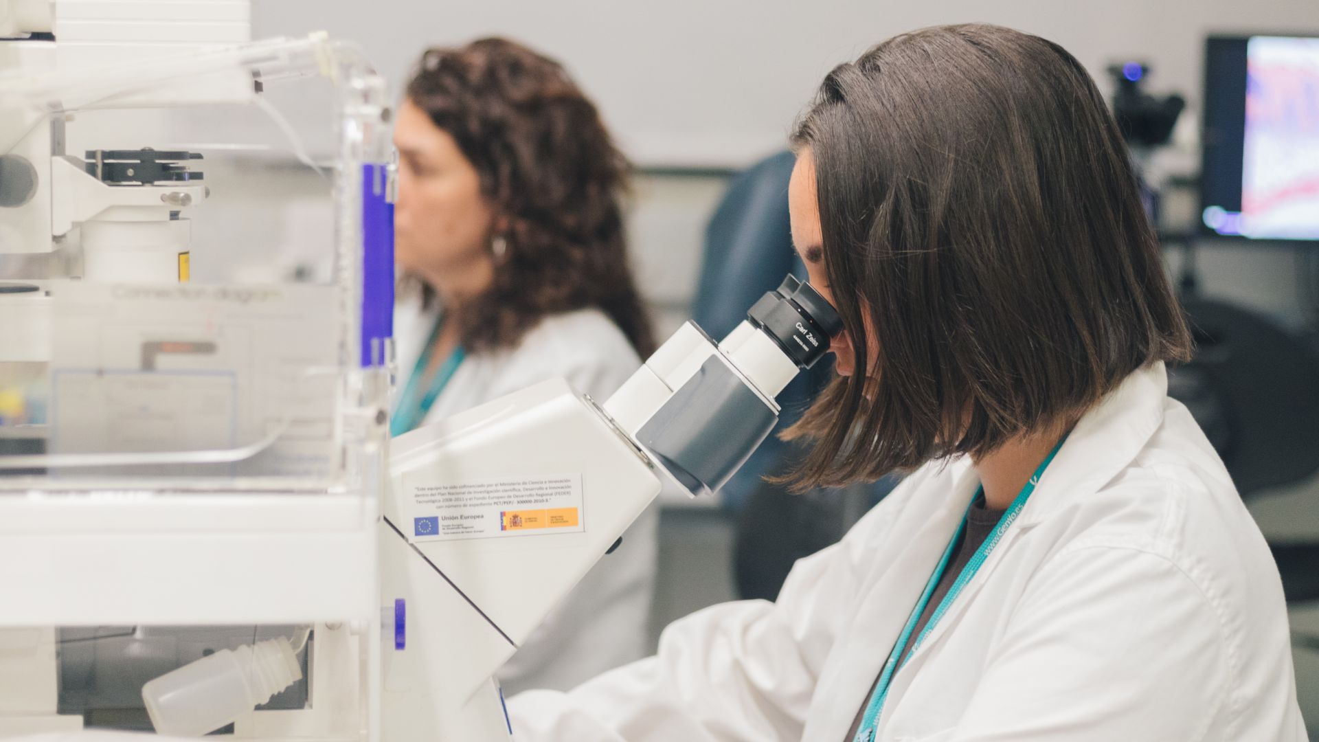
The Microscopy Unit at GENyO is dedicated to the application and development of different advanced confocal laser microscopy techniques, such as the analysis of the localisation and dynamic distribution of molecules of interest in cells or animal models, opening up a wide range of new possibilities for dynamic studies of cellular processes. It also has specific equipment to perform the Total Internal Reflection Fluorescence (TIRF) microscopy technique, in order to visualise processes of cell mobility, protein aggregation and adhesion, endocytosis, exocytosis and vesicle transport, among others, processes located in the basal plasma membrane region of cells. The unit also carries out the technique of microdissection by laser capture, which allows the isolation of pure cell populations or individual cells for subsequent genomic or proteomic study.

Mission

The Microscopy Unit’s main task is to provide technical support in the areas of epifluorescence microscopy, confocal laser microscopy, TIRF microscopy and laser capture microdissection, both to the centre’s research groups and to external entities.

Thanks to the unit’s state-of-the-art technological equipment and the extensive experience of the scientific and technical staff responsible for epifluorescence microscopy, advanced confocal microscopy, TIRF microscopy and laser microdissection, it is possible to develop the applications required by users, and to achieve one of GENyO’s priorities, which is the integration of basic, applied and translational research, minimising the time between the development of new technologies, products and procedures and their application in the healthcare field. As well as participating in the generation of new systems for the diagnosis, prevention and treatment of diseases associated with human genetic variability through the application of new image-based technologies, in order to achieve research excellence in the area of oncology and genomics applied to health.

Members
Scientific Responsible
Technical Responsible

Support Unit Technician

Raquel Marrero

Research support technicians

Support Unit Technician

Alba Pañella

Service Portfolio
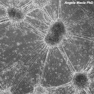
Conventional transmission and epifluorescence microscopy.
  • Image acquisition with transmitted light techniques: brightfield, phase contrast and interferential contrast (DIC).
  • Fluorescence imaging: from fixed samples with a single fluorochrome (single labelling) or multiple fluorochromes (multiple labelling).
Multispectral super-resolution confocal laser microscopy
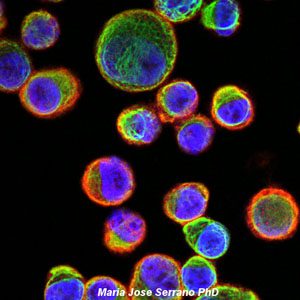
  • Multi-labelling: acquisition of high-resolution, high-sharpness and high-contrast images from samples containing multiple fluorescent markers without signal overlap, through customised detection of the emission spectra of each fluorochrome.
  • Multi-position: acquisition of images from multiple positions (multi-well plate) with high spatial resolution, excellent sensitivity and high experimental reproducibility.
  • Co-localisation: analysis of the overlap in signal distribution in samples containing fluorescent markers. Representation of the degree of co-localisation between fluorescent molecules using: biparametric histograms (a graph showing the fluorescence intensities of the labels of interest plotted against each other), masks (images showing only the pixels that co-localise in both fluorescent labels) and co-localisation coefficients (indices quantifying the level of co-localisation of the fluorescent signals).
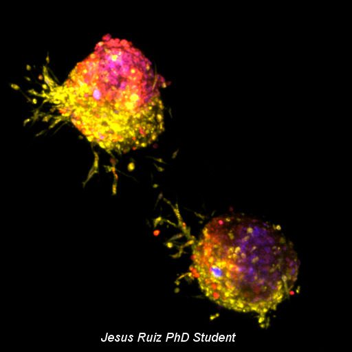
  • Z-Stack: acquisition of optical sections along the Z-axis of the sample and superimposition of the images to generate plane projections that allow 3D reconstructions of the specimen to be visualised.
  • Tile Scan: acquisition of a mosaic image of the sample by capturing and merging multiple images along the XY axes, followed by reconstruction using stitching algorithms, shadow correction and focus adjustment.
  • Time-lapse: the acquisition of a series of images of living samples over time. The equipment features an opaque incubation system that encloses the entire microscope, creating a stable thermal environment that minimises temperature gradients and condensation. It enables precise, automated control of temperature, CO₂ and relative humidity, ensuring the viability of live samples and the stability of the medium’s pH throughout the experiment.
  • Super-resolution: the Airyscan 2 module combines a high-sensitivity multi-pixel detector with advanced reconstruction algorithms to achieve a significant improvement in spatial resolution. The system achieves a lateral resolution of 90 nm and an axial resolution of 350 nm, whilst maintaining high signal capture efficiency and reducing the effects of photobleaching and phototoxicity.
Time Lapse Microscopy Imaging.
  • “In vivo” tests: acquisition of image sequences of live samples with transmitted light and/or fluorescence techniques over time under controlled CO2 and temperature conditions. In addition to the application of multi-position parameters and/or optical sectioning generating XYT or XYZT images.
Photoactivation
  • Photoactivation assays: pre-photoactivation imaging of live samples expressing the molecule of interest fused to a photoactivatable fluorescent protein, selection and activation of a region of interest (ROI) in the sample with the appropriate laser line, and acquisition of an image sequence over time to analyse the dynamic distribution of the fluorescent molecule of interest.
FRAP: Fluorescence Recovery After Photobleaching
  • Fluorescence recovery assays after photobleaching: pre-photobleaching imaging of live samples expressing the molecule of interest fused to a fluorescent protein, selection and photobleaching of a region of interest (ROI) in the sample with the appropriate laser line at maximum power and capture of an image sequence over time to analyse the mobilisation of the molecule of interest by fluorescence recovery in the ROI.
  • Quantification and data analysis: normalisation of results, fluorescence recovery graphs, calculation of the mobile and immobile fraction and the mean fluorescence recovery time.
FLIP: Fluorescence Loss in Photobleaching
  • Fluorescence loss in photobleaching assays: capture of images prior to photobleaching of live samples expressing the molecule of interest fused to a fluorescent protein, selection and continuous photobleaching of a region of interest (ROI) in the sample with the corresponding laser line at maximum power and obtaining a sequence of images over time to analyse the dynamic distribution of the molecule of interest through the generalised loss or decrease of fluorescent molecules in the sample.
  • Quantification and data analysis: normalisation of results and obtaining graphs of fluorescence loss of the molecule of interest.
FRET: Fluorescence Resonance Energy Transfer.
  • Fluorescence resonance energy transfer assays: analysis of intra- or intermolecular interactions, conformational changes or proteolysis processes of the molecule of interest fused to a FRET pair.
  • Advice on sample preparation: selection of the most suitable FRET pairs (donor and acceptor) between fluorescent proteins or secondary antibodies, analysis of co-localisation between donor and acceptor, study of the partial overlap of the donor emission spectrum with the acceptor excitation spectrum and analysis of the lifetime of the fluorescence emitted by the donor.
  • Selection of the FRET detection method:
    • Acceptor Photobleaching method: acquisition of pre-photobleaching images of the donor and acceptor from the FRET sample excited with the corresponding laser lines, selection of a region of interest (ROI) in the acceptor image, continuous photobleaching of the acceptor and subsequent imaging of the donor and acceptor excited with the corresponding laser lines.
    • Sensitised Emission method: capture of reference images of the donor and acceptor from the control samples with the corresponding wavelength laser lines, acquisition of donor and acceptor images in the FRET sample with the donor excitation laser line, and finally, acquisition of only the acceptor image in the FRET sample with the acceptor excitation laser line.
  • Data quantification and analysis: normalisation of results, obtaining plots of donor and acceptor fluorescence intensity, and pixel-by-pixel calculation encoded in a pseudo-coloured image showing the % FRET efficiency.
TIRF: Total Internal Reflection Fluorescence Microscopy
  • Total Internal Reflection Fluorescence Fluorescence (TIRF) microscopy assays: acquisition of fluorescence images of fixed or living cells, by selective excitation of fluorophores located in the region adjacent to the sample/glass interface with the appropriate laser line (488 nm and/or 561 nm) and detection of the emitted signal with a high-sensitivity, low-noise monochrome EM-CCD camera, under controlled CO2 and temperature conditions in the case of living specimens, to analyse dynamic processes of cell motility, protein aggregation and adhesion, endocytosis, exocytosis, vesicular trafficking, which take place in the contact zone between the basal plasma membrane of the cells and the glass or substrate to which they adhere.
Microdissection and laser catapulting
  • Fixed cells: UV laser catapulting of immunohistochemically or immunofluorescently labelled cells.
  • Tissue sections: microdissection and UV laser catapulting of regions of interest from cryo-frozen or FFPE tissue sections, labelled with immunohistochemistry or immunofluorescence techniques for subsequent DNA (PCR, mutation analysis, SNPs…), RNA (qRT-PCR, expression analysis, microarrays…) or protein (immunoblot, 2D gels, MALDI-TOF…) extraction assays.
Nanoparticle tracking analysis (NTA)
  • Tracking of bio-nanoparticles (extracellular vesicles, exosomes, liposomes, protein aggregates, viruses) and synthetic nanoparticles suspended in polar liquids and organic solvents for studies of size, concentration, fluorescence and zeta potential, through real-time visualisation of Brownian motion and electrophoretic mobility. Up to four fluorescence channels with independent excitation and emission are available. A co-localisation function is available between two fluorescence channels of interest.
Technical advice on experimental design
  • The unit provides technical support in the design of experiments and the correct preparation of samples.
Technical support in image processing and analysis
  • We offer technical support for image processing and analysis using various software programmes (including Arivis Pro 4.4.0, Zen 3.13, PALMRobo, NIS-Elements and Fiji, amongst others), as well as for the correct interpretation and presentation of the image data obtained.
Supply of consumables for "in vivo" assays

Confocal Microscopy:

  • IBIDI 4/8-well plates
  • IBIDI 50 mm plates with bottom coverslip
  • IBIDI 35mm plates with bottom coverslip

Microdissection and laser catapulting:

  • PEN membrane slides
  • PET membrane slides
  • Opaque adhesive collector tubes 500 µl
  • Opaque adhesive collector tubes 200 µl
  • Transparent adhesive collector tubes 200 µl
Equipment
Epifluorescence microscopes
  • Nikon Eclipse 50i microscope: Upright microscope with 10x, 20x, 40x (dry) and 100x (oil immersion) objectives, transmitted light techniques (brightfield, phase contrast and DIC) and filters (DAPI, FITC and TxRed).
  • Nikon Eclipse TE2000-U microscope: Inverted microscope with 4x, 10x, 20x, 40x and 60x objectives (dry), transmitted light techniques (brightfield and phase contrast) and filters (DAPI, FITC and TxRed).
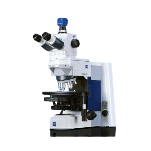
  • Zeiss Axio Imager A.1 microscope: Upright microscope with 5x, 10x, 20x (dry), 63x and 100x (oil immersion) objectives, transmitted light techniques (brightfield and phase contrast) and filters (BFP, DAPI, A488 and Rhod).
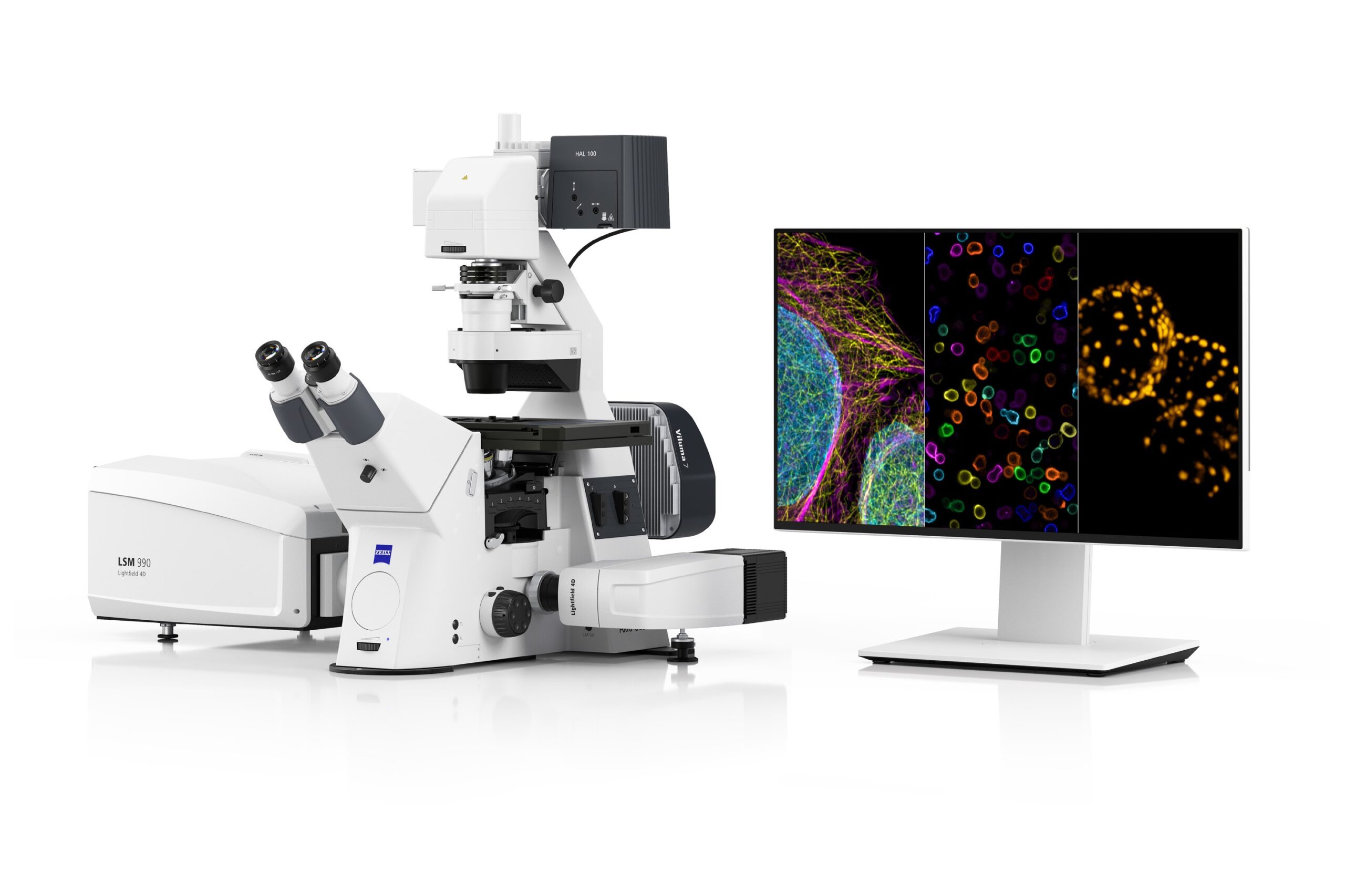
ZEISS LSM 990 AIRYSCAN 2 confocal laser microscope
  • Inverted confocal laser microscope with 7 diode laser excitation lines (405, 488, 514, 561, 594, 639 and 730 nm), 5 objectives (5x dry AN 0.16, 10x dry AN 0.45, 25x multi-immersion AN 0.8, 40x multi-immersion AN 1.2 and 63x oil immersion AN 1.4), transmitted light techniques (brightfield and DIC), 4 fluorescence filters (DAPI, GFP, DsRed and Cy5), 7 detectors (2 lateral PMTs, 1 central GaAsP, 1 Airyscan, 2 NIR (Near-Infrared) and 1 T-PMT), fully motorised and equipped with an incubation system featuring CO₂, temperature and humidity control for experiments with live samples.
  • ‘Airyscan 2’ super-resolution module based on a monochromatic GaAsP detector array comprising 32 optical elements that act collectively as a single specialised, high-sensitivity detector. The system achieves a lateral resolution of 90 nm and an axial resolution of 350 nm. The detector allows up to 4 pixel lines to be recorded simultaneously in parallel, achieving speeds of up to 25 fps at 512 × 512 pixels; for live samples, it can scan up to 8 lines simultaneously, achieving speeds of 47.5 fps at 512 × 512 pixels.
  • The innovative “Sample Finder” tool automates the localisation and centring of samples, providing an overview of the sample holder or plate and allowing regions of interest to be selected intuitively via the software. It facilitates navigation between multiple acquisition areas, which is particularly useful in screening, multi-well or mosaic experiments.
  • The “Definite Focus 3” hardware-based autofocus system, utilising an infrared laser, ensures continuous and precise focal stability during long-running experiments or automated acquisitions. It compensates for deviations caused by temperature changes, vibrations or sample irregularities, guaranteeing consistent image quality and high reproducibility, particularly in long-running experiments, mosaic acquisitions, multi-well plate analyses and experiments with live samples.
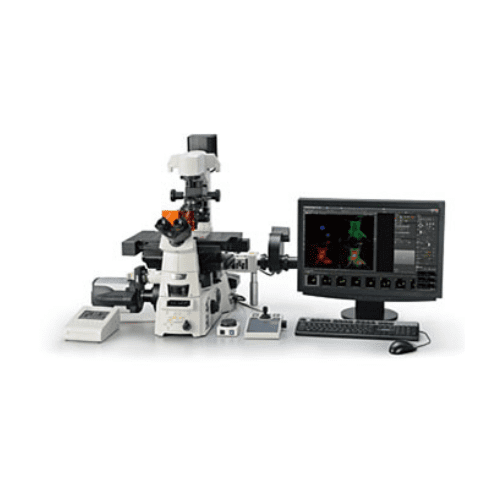
Epifluorescence microscope with TIRF module Nikon Eclipse Ti-E
  • Epifluorescence microscope with inverted TIRF module with 4 excitation laser lines: Diode (405 nm), Argon (457, 477, 488 and 514 nm), Diode (561 nm) and Diode (638 nm), objectives 10x, 40x (dry), 100x TIRF (oil immersion), transmitted light techniques (brightfield and phase contrast), filters (DAPI, YFP, G2-A and TIRF GFP/RFP), with high-sensitivity monochrome EM-CCD Andor iXON DU885 camera, fully motorised, integrated Perfect Focus System (PFS) and incubation cabinet with CO2 and temperature control for live sample testing.
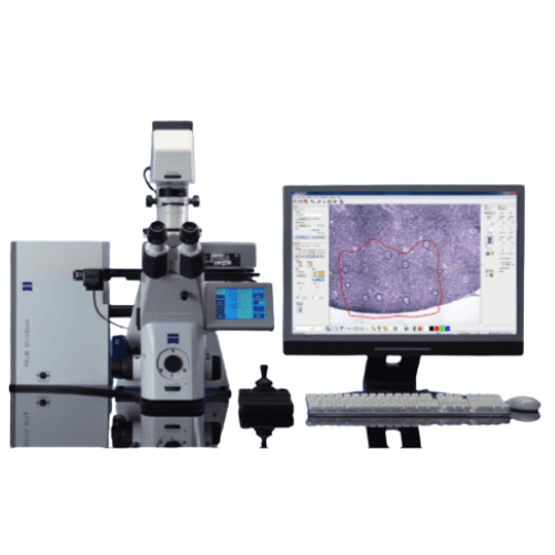
Zeiss PALM Microbeam IV Laser Microdissector Zeiss Laser Microbeam IV
  • Inverted laser microdissector with 1 excitation laser line in the UV range (355 nm), 10x, 20x, 40x and 63x objectives (dry), transmitted light techniques (brightfield, phase contrast and DIC), filters (DAPI, GFP and Rhod), fully motorised, high precision in cutting and catapulting and with ‘cap check’ system to check the presence of the catapulted sample in the adhesive cap of the collecting tube.
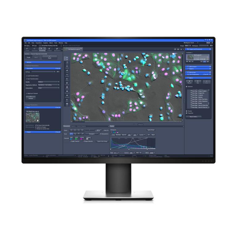
Image acquisition, processing and analysis software


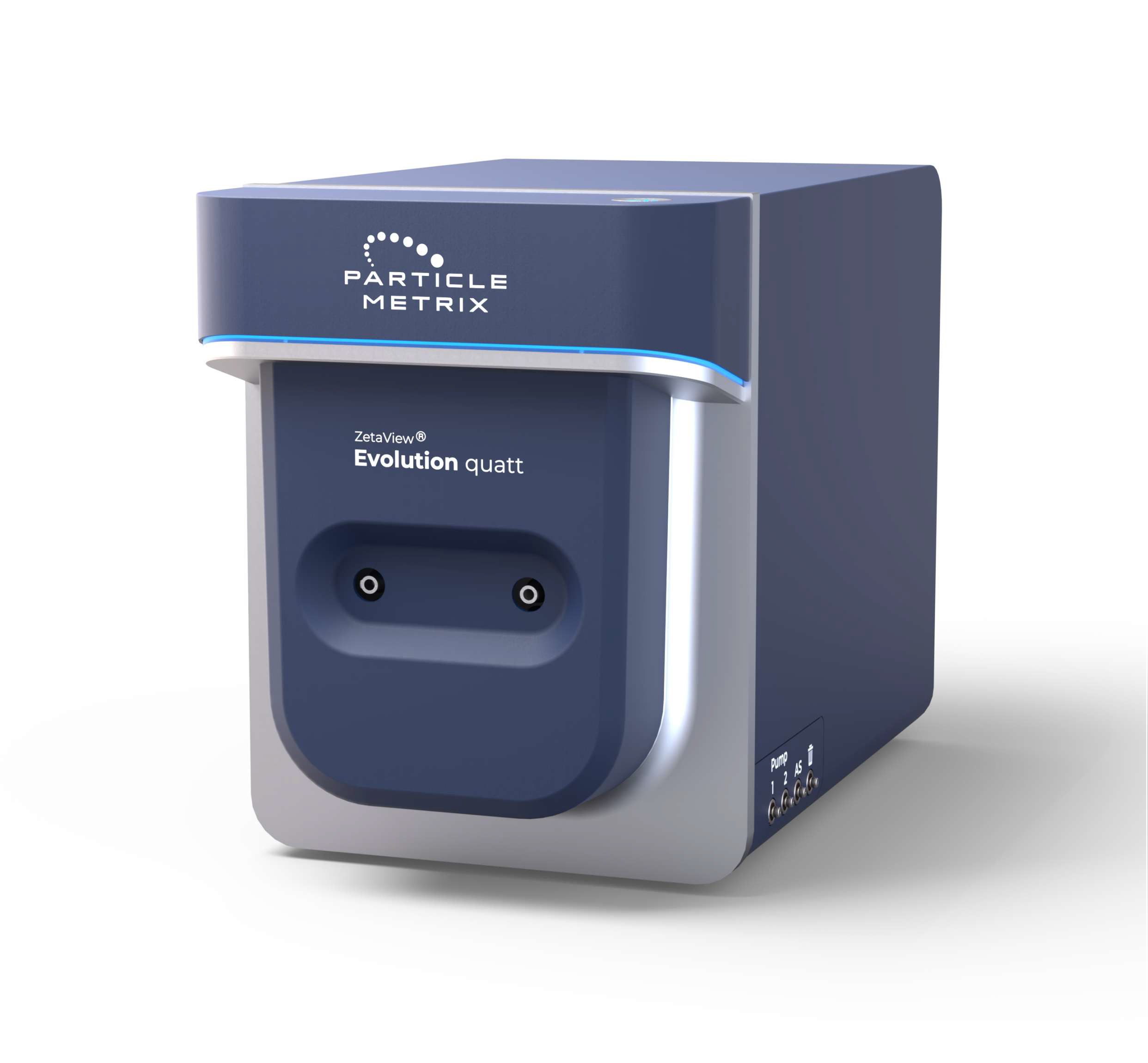
PARTICLE METRIX ZETAVIEW EVOLUTION QUATT nanoparticle tracking analysis (NTA) system

Motorised scanning nanoparticle tracking analysis (NTA) instrument for tracking the movement of individual nanoparticles in suspension. It features an 11-position, software-controlled fluorescence emission filter wheel for rapid switching between scattering and fluorescence measurements, as well as between different emission filters. It has 4 fluorescence channels. Available lasers and filters:

  • 405 nm laser. Cut-off filter at 410 nm.
  • 488 nm laser. Cut-off filter at 500 nm.
  • 520 nm laser. Cut-off filter at 550 nm.
  • 640 nm laser. Cutoff filter at 660 nm.
Galery

Subscribe to our newsletter