Microscopy and Molecular Imaging Unit

Home Support units Microscopy and Molecular Imaging Unit

Microscopy and Molecular Imaging Unit

Microscopy and Molecular Imaging Unit. Introduction

Optical microscopy is a technique use to examine specimens mainly transparent. Opaque specimens are considered if the reflection surface is not completely pulished. Light passes through the specimen at different depths, which causes a blurry image due to the light detection coming from areas out of the plane of focus, reducing importantly sharpness, contrast, and image resolution.

Confocal laser microscopy technique became very successful after the development of laser technique. It obtained excellent results in several areas such as medicine, biology, or geology, because of its unquestionable advantages compared to the conventional optical microscopy. The principle of confocal laser microscopy is based on the elimination of the reflected light or fluorescence from the planes out of focus. To that end, the specimen is illuminated point by point with a laser line, and the only light detected is that of the focal plane, then, eliminating the light beams above and below the plane of focus, thus images of high sharpness, contrast and resolution are created. Also, confocal laser microscopy enables to obtain optical sections of the specimen, which allows the study of 3D images.

Laser capture microdissection is focused on the observation and separation of unique cells or small cell groups from a heterogeneous tissue in an efficient, rapid and targeted manner, which makes possible the systematic study of the quality and quantity of biomolecules (DNA, RNA, or proteins) in specific cell populations. This technology, firstly developed for molecular analysis of tumors, can be applied on a wide variety of tissues, and facilitates the study of the molecular bases of many diseases such as cancer, rare diseases, degenerative diseases, diabetes, and hypertension, among others.

Microscopy and Molecular Imaging Unit in GENYO is focused on the application and development of different advanced techniques of confocal laser microscopy, as well as the analysis of the dynamic localization and distribution of molecules of interest in cells or animal models, opening a wide range of new possibilities in addressing dynamic studies of cellular processes. Furthermore, laser capture microdissection technique available in the Unit, which allows isolation of pure cell populations or individual cells to the subsequent genomic or proteomic study.


Mission

The main task of the Microscopy and Molecular Imaging Unit is to provide technical support in the area of confocal laser microscopy and laser capture microdissection, both to the research groups of the Center and outside entities.

The next generation technological equipment of GENYO, together with the large experience of the scientific and technical team specialized in advanced confocal microscopy and laser microdissection, makes it possible to develop the applications required for the users of this Unit, and to achieve the main priorities of GENYO, such as the integration of basic, applied, and translational research, minimizing the times between the development of new technologies, products and procedures, and their application in the health setting. The Unit also makes it possible to participate in the development of new diagnostic systems, prevention and treatment of diseases associated to human genetic variability, by the application of new imaging techniques, in order to reach an investigation of excellence in oncology and genomics areas applied to the health care.

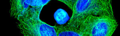
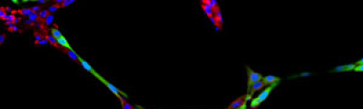
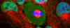
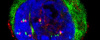
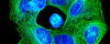
The activity of the Microscopy and Molecular Imaging Unit is focused on epifluorescence microscopy and confocal laser microscopy, which allows to analyze the localization of molecules of interest using immunofluorescence techniques and fluorescent fusion proteins such as CFP, GFP, YFP, mRFP, mCherry, among others, both in fixed and living cells, which makes it possible to develop studies on dynamic cellular processes, among others. Furthermore, another activity of the Unit is laser capture microdissection technique, which facilitates the isolation of specific cell populations in heterogeneous tissue sections or individual cells for subsequent genomic and proteomic study.

Epifluorescence Microscopy

The light source in epifluorescence microscopy is a lamp with light containing different wavelengths. A filter is used to gain the adequate excitation wavelengths, depending on the fluorochrome of the specimen, so that the reflected light or emitted fluorescence light after passing through an emission filter system, reaches the detector or camera, then obtaining a digital image in the computer.

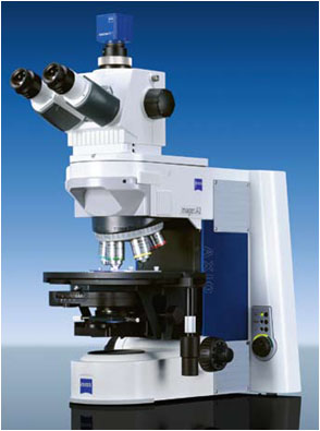
The Microscopy and Molecular Imaging Unit of GENYO is provided with the following epifluorescence microscopy equipment : Nikon Eclipse 50i, Nikon Eclipse 80i, Nikon Eclipse 90i with spectral camera and motorized plate, Nikon Eclipse TE2000-U, and Zeiss Axio Imager A.1, providing images acquisition with transmitted-light techniques (bright field, phase contrast, and DIC), and fluorescence images acquisition from fixed specimens with one or several fluorochromes (single or multiple labeling, respectively).

Confocal Laser Microscopy

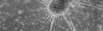
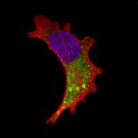
In contrast to epifluorescence microscopy, confocal laser microscopy allows to obtain high-resolution images, as well as sharpness, and contrast of only one focal plane of the specimen when light from planes out of focus is eliminated. Confocal laser microscope captured images of the specimen that reflect fluorescent light; in that case, the light source is a laser line, which excites the specimen point by point and moves along the focal plane by a laser scanning system.

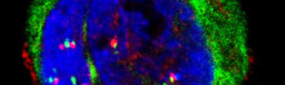
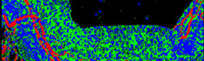
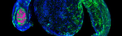
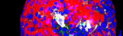
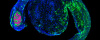
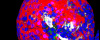
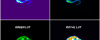
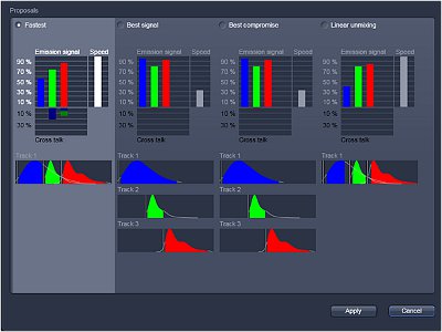
Fluorescence from the focal plane of the specimen is detected by a photomultiplier tube, and converted into electrical signal, which is digitalized and converted into a signal for display into a video monitor, resulting in an exceptional quality image. Confocal laser microscope allows the study of specimens marked with several fluorochromes, by the capture, without overlap, of the signals coming from the fluorochromes through the tailor-made detection of the emission spectra of each fluorochrome; Then, multiple images are generated, one for every fluorochrome detected, which can be finally overlapped in one image.

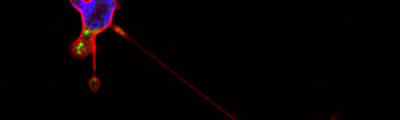
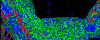
One of the main advantages of confocal microscopy is the possibility of changing the focal plane position, and capturing images at different Z-axis heights of the specimen. Then, obtaining a set of optical sections to which it is possible to apply reconstruction techniques able to provide visualization of 3D structure of the specimen.

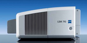
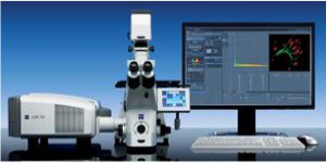
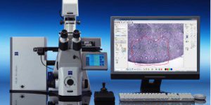
GENYO is equipped with the last generation technology of confocal laser microscopy such as the LSM 710 (Zeiss) confocal laser microscope, equipped with an incubation chamber with temperature and CO2 control to perform assays with living specimens.

Laser Capture Microdissection

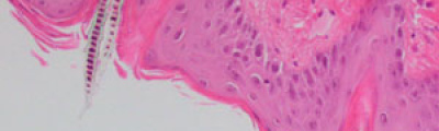
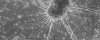
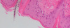
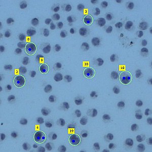
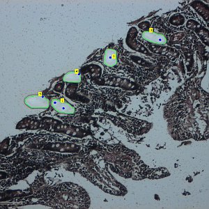
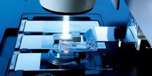
Laser Capture Microdissection allows the isolation of regions of interest from cryo frozen tissue sections or FFPE tissues, cell populations or even specific individual cells from fixed or living specimens marked with immunohistochemical, immunofluorescence methods, or techniques expressing molecules of interest fused to fluorescent proteins in order to perform a later genomic or proteomic study.

The impossibility of isolating pure groups of cells was a major constraint in the study of a specific type of cell. However, the development and evolution of microdissection techniques resulted in laser capture microdissection system. It is an efficient, accurate and safe from environmental contamination technique, addressed to investigators interested in separating cell populations or specific unique cells for the later molecular analysis.

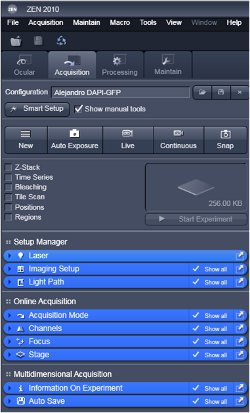
The Microscopy and Molecular Imaging Unit of GENYO is equipped with the Zeiss PALM Microbeam IV Laser capture microdissection system. The Zeiss Axio Observer Z.1 inverted microscope is available for tissue or cellular sections observation providing excellent quality images. Thanks to the PALMRobo software, tissue or cellular regions of interest can be easily and rapidly localized on the screen. The drawing tools of the program allow toselect cellular groups or unique cells. The system’s UV laser (355 nm) is used to both cut and catapult cellular groups or unique cells into the adhesive cap of the collection tube to be studied. The “Cap Check” system of the program is used to verify the catapulted sample in the cap, which provide a higher reliability of the system.

The Unit is equipped with Epi-fluorescence microscopy, confocal laser microscopy, and laser capture microdissection systems. The main characteristics of all of them are the following:

Epi-fluorescence Microscopes:

  • Microscopio Nikon Eclipse 50i:Upright microscope with 10x, 20x, 40x (dry objectives) and 100x (oil immersion objectives), transmitted light techniques (bright field, phase contrast, and DIC) and DAPI, FITC y TxRed filters.
  • Microscopio Nikon Eclipse 80i:Upright microscope with 10x, 40x (dry objectives) and 100x (oil immersion objectives), transmitted light techniques (bright field, and DIC) and DAPI, FITC, TxRed, FITC/TxRed, Sp.GOLD, and Sp.AQUA filters.
  • Microscopio Nikon Eclipse 90i ProScanIII: Upright microscope with spectral camera 420-720 nm, and 2x, 10x, 20x y 40x dry objectives, transmitted light techniques (bright field, and DIC) and DAPI, FITC, E-94 (orange) and E-93 (red) fully motorized.
  • Microscopio Nikon Eclipse TE2000-U,Inverted microscope with 4x, 10x,20x,40x, and 60x objectives (dry-type), transmitted light techniques (bright field, and phase contrast) and DAPI, FITC and TxRed filters.
  • Microscopio Zeiss Axio Imager A.1:
    Upright microscope with 5x, 10x, 20x (dry objectives), 63x, and 100x (oil immersion objectives), transmitted light techniques (bright field, and phase contrast) and BFP, DAPI, A488, and Rhod filters.

Confocal Laser Microscope

    • Microscopio Láser Confocal Inverted microscope with 5 excitation laser lines: diode laser (405 nm), Argon laser (458 nm, 488 nm, 514 nm), HeNe laser (543 nm, 594 nm, and 633 nm); with 10x, 20x dry objectives, 40x, 63x oil immersion objectives,63x water and glycerol immersion, transmitted light techniques (bright field, and DIC), Zeiss filters for DAPI, GFP, DsRed, and FITC-TRITC, with 3 internal or photomultiplier detection channels (PMTs); the system is fully motorized, with CO2 and temperature controlled incubation system for “in vivo” assays.

Microscopio Epifluorescencia con módulo TIRF Nikon Eclipse Ti-E

    • Microscopio Epifluorescencia con módulo TIRF invertido con 4 líneas de láser de excitación: Diodo (405 nm), Argón (457, 477, 488 y 514 nm), Diodo (561 nm) y Diodo (638 nm), objetivos 10x, 40x (en seco), 100x TIRF (inmersión en aceite), técnicas de luz transmitida (campo claro y contraste de fases), filtros (DAPI, YFP, G2-A y TIRF GFP/RFP), con cámara monocroma de alta sensibilidad EM-CCD Andor iXON DU885, completamente motorizado, sistema de enfoque perfecto integrado (PFS) y cabina de incubación con control de CO2 y temperatura para realizar ensayos con muestras vivas.

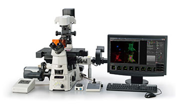
Zeiss PALM MicroBeam IV Laser Microdissection instrument

    • Microdisector Láser with one excitation laser line of ultraviolet range (355nm), objectives 10x,20x,40x and 63x (dry-type), transmitted light techniques (bright field, phase contrast, and DIC); DAPI, GFP and Rhod filter set; the system is fully motorized, with high precision cutting and catapulting, and “cap check” system.

Processing Software and Imaging Analysis

List of Services

01Conventional Transmission Microscopy and Epi-fluorescence Microscopy
    • Image Acquisition using transmitted light techniques: bright field, phase contrast, and DIC..
    • Fluorescence Images Acquisition: From fixed specimens with one or several fluorochromes (single or multiple labeling, respectively).
02Multispectral laser confocal microscopy
    • Multiple labeling: acquisition of high-resolution, sharpness and contrast images from fixed specimens, presenting multiple fluorescent markers without overlap of signals through tailor-made detection of fluorochromes emission spectra.
    • Co-localization: It refers to the overlap analysis of the distribution of signals in fluorescent markers specimens, and represents the colocalization degree between fluorescent molecules using biparametric histograms (diagram showing different fluorescence intensities of labeled species of interest ), mask (image of colocalized pixels in both fluorescent labelings), and colocalization coefficient (quantification of the degree of colocalized fluorescent signals).
    • Z-Stack: Obtention of optical sectioning in the Z-axis of the specimen, and images overlapping, then producing maximum intensity projections (MIP) of planes that allow visualization of 3D specimen reconstruction.
    • Tile Scan: acquisition of a mosaic image of the specimen by capture of multiple images in the XY axes.
03Time Lapse Microscopy Imaging.
    • “In vivo” assays: acquisition of imaging sequences of live specimens with over time transmitted light and/or fluorescence techniques in controlled Co2 and temperature conditions. Also, application of multi-positions parameters and/or optical sectioning, then generating XYT or ZYZT images.
04Photo-activation
    • Photo-activation Assays: Capture of images before photoactivation of live specimens that express the molecule of interest fused to a photoactivatable fluorescent protein, selection and activation of a region of interest (ROI) in the specimen with the corresponding laser line, and acquisition of an images’ sequence over time to analyze the dynamic distribution of the fluorescent molecule of interest.
05FRAP: Fluorescence Recovery After Photobleaching.
    • Fluorescence Recovery After Photobleaching Assays: Obtaining images before photobleaching of live specimens that express the molecule of interest fused to a fluorescent protein, selection and photobleaching of a region of interest (ROI) in the specimen with the corresponding full power laser line, and obtention of an image sequence over time to analyze mobilization of molecules of interest by fluorescence recovery in the ROI.
    • Data quantification and analysis: normalization of results, recovery fluorescence graphics, calculation of the mobile/immobile fraction and half-time fluorescence recovery.
06FLIP: Fluorescence Loss in Photobleaching.
    • Fluorescence Loss in Photobleaching Assays: capture of images before photobleaching of living specimens that express the molecule of interest fused to a fluorescence protein, selection and continuous photobleaching of a ROI in the specimen with the corresponding full power laser line, obtention of an images’ sequence over time to analyze the dynamic distribution of the molecule of interest by the loss or overall reduction of fluorescent molecules in the specimen.
    • Data quantification and analysis: normalization of results, obtention of graphics about fluorescence loss of the molecule of interest.
07FRET: Fluorescence Resonance Energy Transfer.
    • Fluorescence Resonance Energy Transfer: analysis of intramolecular or intermolecular interactions, conformational changes or proteolysis process of the molecule of interest fused to a FRET pair.
    • Technical advice in the sample preparation: selection of the most adequate FRET donor/acceptor pairs between fluorescent proteins or secondary antibodies, analysis of the co-localization between donor and acceptors, study of the partial overlap of the emission spectrum of the donor with the excitation spectrum of the acceptor, and measurement of the donor fluorescence lifetime.
    • Selection of the FRET detection method:
  • FRET Acceptor Photobleaching method: acquisition of images before donor and acceptor photobleaching of the FRET sample, excited with laser lines; selection of a ROI in the acceptor image; continuous acceptor photobleaching and the later capture of images of the donor and acceptor excited with laser lines.
  • Sensitized Emission Method: capture of reference images of donor and acceptor from control samples with the corresponding wavelength laser lines; images acquisition of the donor and acceptor in the FRET sample with donor excitation laser line, and finally, image acquisition of the acceptor alone in the FRET sample with the acceptor excitation laser line.
    • Data quantification and analysis: normalization of results, obtention of graphics of the donor and acceptor fluorescence intensity, pixel by pixel calculation codified in a pseudocolored image relative to the percentage of FRET efficiency.
08Microscopía TIRF: Total Internal Reflection Fluorescence.
    • Ensayos de microscopía de Fluorescencia de Reflexión Interna Total (TIRF): adquisición de imágenes de fluorescencia de células fijadas o vivas, mediante excitación selectiva de fluoróforos localizados en la región adyacente a la interfase muestra/vidrio con la línea de láser adecuada (488 nm y/ó 561 nm) y detección de la señal emitida con cámara EM-CCD monocroma de alta sensibilidad y bajo ruido, en condiciones controladas de CO2 y temperatura en el caso de especímenes vivos, para analizar procesos dinámicos de movilidad celular, agregación y adhesión de proteínas, endocitosis, exocitosis, tráfico vesicular, que se desarrollan en la zona de contacto entre la membrana plasmática basal de las células y el vidrio o sustrato sobre el que se adhieren.
09Laser Microdissection and Catapulting
    • Fixed cells: UV laser catapulting of cells marked with immunohistochemical or immunofluorescent techniques.
    • Tissue Sections: UV laser microdissection and catapulting in ROI from cryo-forzen tissue sections or FFPE, marked with immunohistochemical or immunofluorescent techniques to be used for later extraction assays of DNA (PCR, mutation analysis, SPs…), RNA (qRT-PCR, expression analysis, microarrays…) or protein (immunoblotting, 2D gels, MALDI-TOF…).
10Technical Advice on the experiment design
    • The Unit provides technical advice and assessment on the design of experiments and the correct preparation of the samples.
11Technical support on the images processing and analysis
    • We provide technical support on the processing and analysis of images by using different software programs such as Zen, AxioVision, PALMRobo, MetaMorph, NIS-Elements, Image J, and Adobe Photoshop, as well as on the correct interpretation and presentation of the data of the images obtained.
12Supply of consumable material for “in vivo” assays of confocal laser microscopy and laser capture microdissection
    • Confocal Microscopy:
    • Laser Microdissection and catapulting:

The Microscopy and Molecular Imaging Unit is under the scientific direction of Professor. Dr. María José Serrano Fernández, PhD. who has large experience in the field of confocal laser microscopy and laser capture microdissection.

Dr. Serrano Fernández obtained her PhD degree in 2003 at the University of Jaén, Spain. She worked at the Health Sciences Department., and focused her investigation on the study of the phenotypic and genetic characteristics of circulating tumor cells present in the blood of solid tumor patients. These studies imply the use of different microscopy systems together with phenotypic and genetic analysis software. A patent has been developed under this line of research based on the phenotypic analysis of circulating tumor cells with multiple markers. She is completing the studies by using platforms such as laser microdissection systems, since laser capture microdissection is a useful and reliable technique in the isolation of pathologic pure cell populations into the tumor mass. This method together with molecular biology and confocal laser microscopy, is to distinguish which molecules are involved in the metastatic process and the tumor dissemination.


Dr. Raquel Marrero Díaz is the responsible for the technical direction. She obtained her PhD degree at the Universidad Complutense of Madrid in 2009. She wrote the thesis at the Confocal Microscopy Unit of the Centro Nacional de Investigaciones Oncológicas , where she acquired wide formation and excellent experience in advanced epi-fluorescence microscopy and confocal laser microscopy techniques such as photoactivation, FRAP, FLIP, FRET, time-lapse microscopy imaging, as well as imaging processing and analysis. She is actually tasked with managing and maintaining the microscopy and laser microdissection equipments of GENYO. She also provides technical support to internal and external users of the Unit, on the experiments design and the correct samples preparation. She also conducts training courses to the research staff about microscopy techniques and processing systems and imaging analysis. She carries out new techniques and advanced applications in cooperation with the scientific director in order to improve quality in services of the Unit.

Contact

If you are interested in any service of the Microscopy and Molecular Imaging Unit, you have to implement the Application Form to register as new user, and send it to one of the following addresses: